
Kazuo Yamada MD Background and PurposeB-mode ultrasonography has been used to evaluate cervical carotid artery lesions. The HeartFlow Analysis is the first and only non-invasive test which enables your physician to understand the impact that narrowings and blockages have on blood flow to your heart information that otherwise would only be available with a riskier invasive procedure.
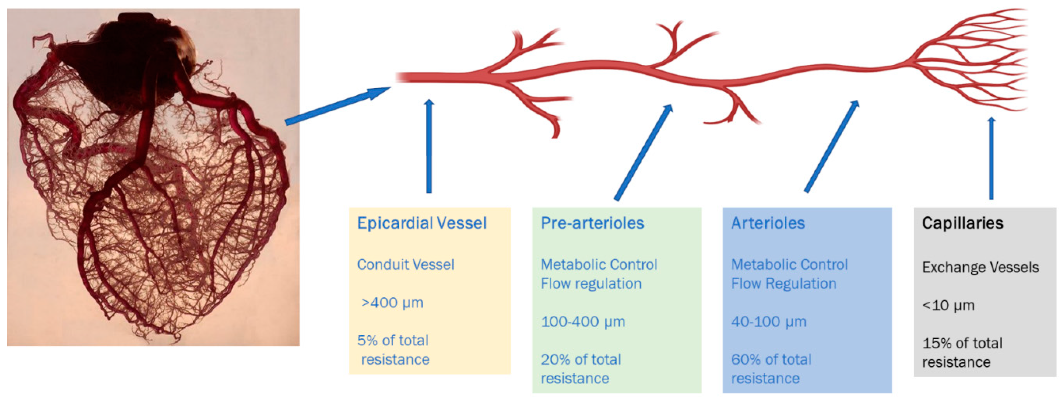
Diagnostics Free Full Text Coronary Microvascular Dysfunction And The Role Of Noninvasive Cardiovascular Imaging Html
B-Mode Flow Imaging of the Carotid Artery Atsushi Umemura MD.

. Since the ultrasound beam is identical to the one used in standard IVUS the colour information is displayed simultaneously with the standard IVUS information. 10 Reliability problems can arise when blood flow monitoring is performed in small ocular arteries or in veins because the lower velocities found in these. Myocardial perfusion scanning is performed using single photon emission computed tomography.
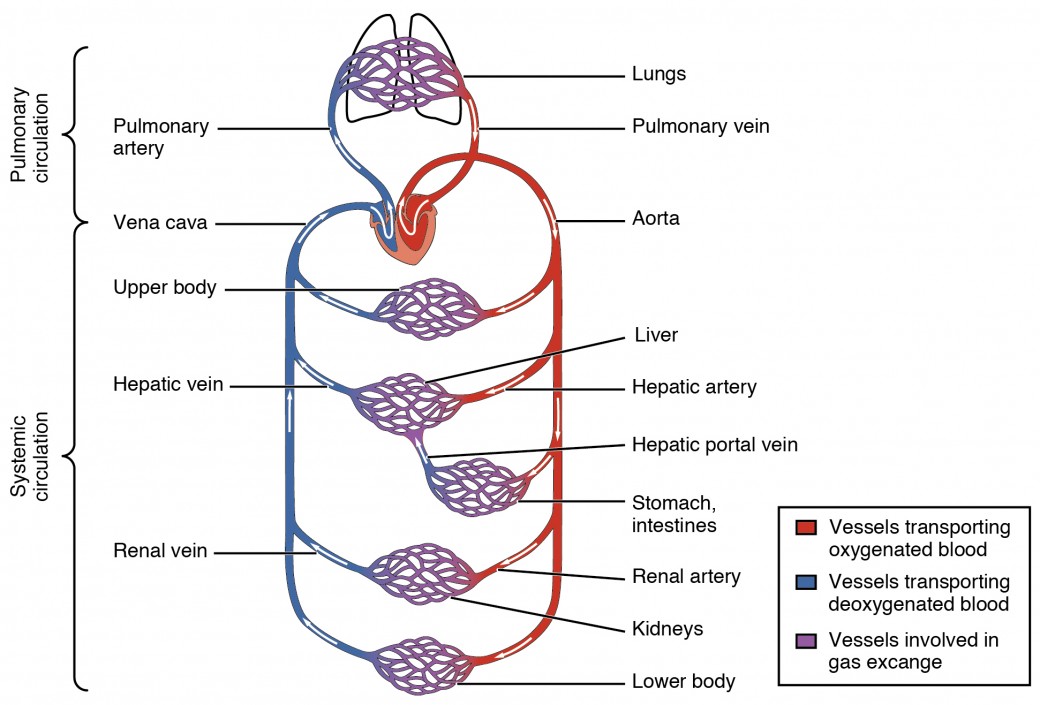
Blood flows through the heart in the following order. Recently a more time. PH-associated vortical blood flow which is typically observed rotating in a.
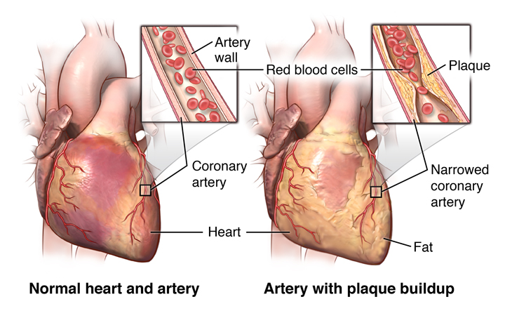
Technologies that are used to generate images of the blood vessels include. Radionuclide imaging is also used to assess improvement in blood supply to the heart muscle after bypass surgery Coronary Artery Bypass Grafting Coronary artery disease is a condition in which the blood supply to the heart muscle is partially or completely blocked. Bringing together human ingenuity and advanced technology to help combat heart disease the 1 cause of death.
1 body 2 inferiorsuperior vena cava 3 right atrium 4 tricuspid valve 5 right ventricle 6 pulmonary arteries 7 lungs 8 pulmonary veins 9 left atrium 10 mitral or bicuspid valve 11 left ventricle 12 aortic valve 13 aorta 14 body. The ophthalmic artery is easily visualized deep in the orbital cavity in the area where it crosses the optic nerve. Some reports suggested that abnormal blood flow parameters.
Vascular imaging is used to evaluate blood vessels with the exception of the coronary arteries which are assessed with a CT scan and help diagnose conditions associated with abnormal blood flow. A percutaneous coronary intervention PCI optimization system was used in this study which combines wireless FFR measurement and FD-OCT imaging in one platform. Color Doppler Duplex provides flow information within a region of interest ROI while spectral Doppler provides information within a sample volume.
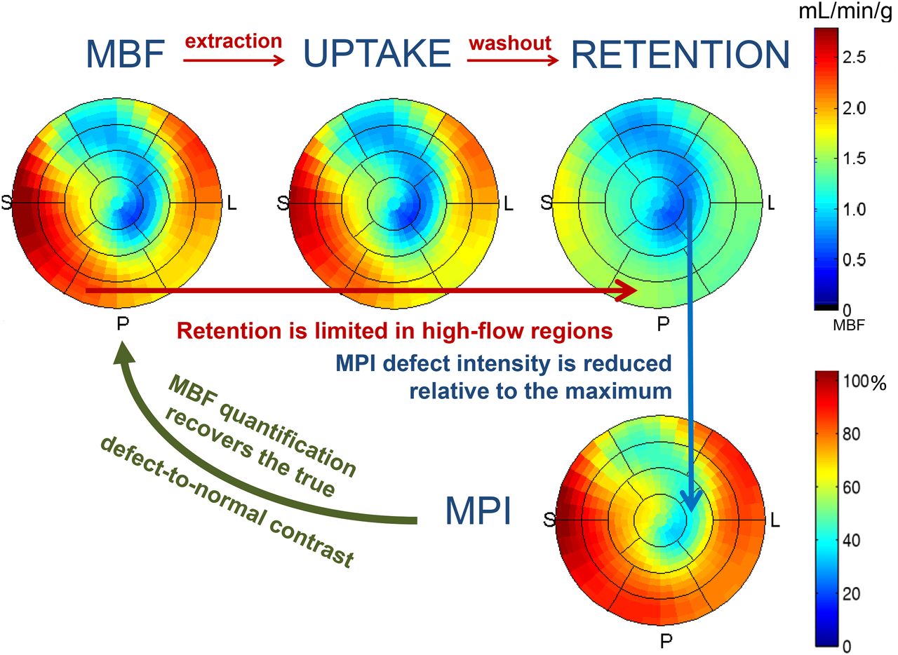
Cardiac imaging by means of myocardial Positron Emission TomographyComputed Tomography PETCT is being used increasingly to assess coronary artery disease to guide revascularization decisions with more accuracy and it allows robust quantitative analysis of both regional myocardial blood flow MBF and myocardial flow reserve MFR. This is important for exploring hemodynamic mechanisms behind vascular diseases associated with arterial pulsations. Recently the technology for direct visualization of blood reflectors has made B-mode flow imaging B-flow possible without the limitations of Doppler technology.
HeartFlows non-invasive personalized cardiac test provides unprecedented visualization of each patients coronary arteries enabling physicians to. Because rapidly moving targets such as red blood cells produce a unique set of echoes in effect one can visualize blood flow patterns this is. We evaluated the efficacy of B-flow in.
Imaging stress tests can lead. The FFR value was 080 in 5 stenoses 25. Doppler is primarily used in longitudinal views but can also useful in a transverse view.
Vascular ultrasound computed tomography CT and magnetic resonance imaging MRI. The test can help your doctor find out if blood flow to your heart is blocked. It can help you and your doctor make decisions about treatment.
Stress may be induced by exercise on a treadmill or by the use of pharmaceuticals such as regadenoson. The mean blood flow rate. The best reason to have an imaging stress test is to manage severe heart disease.
Recently time-resolved 3D phase contrast magnetic resonance imaging 4D-flow allows flow dynamics in patients with pulmonary arterial hypertension to be measured. Blood flow imaging of the fetal heart and great vessels is extremely challenging because the heart beats rapidly the vessels are small and the fetus. It enables visualizing and quantifying blood flow by analyzing interactions of acoustic pulses with flowing blood within the vessel lumen.
If you are at low risk and dont have symptoms the test isnt very useful. If the coronary arteries are narrowed radionuclide imaging is used to learn how the narrowing is affecting the hearts blood supply and function. Magnetic resonance imaging MRI is a noninvasive test that uses a magnetic field and radiofrequency waves to create detailed pictures of organs and structures inside your body.
During a Doppler ultrasound a technician trained in ultrasound imaging sonographer presses a small hand-held device transducer about the size of a bar of soap against your skin over the area of your body being examined moving from one area to another as necessary. The present study aimed to investigate the association between myocardial blood flow MBF quantified by dynamic CT myocardial perfusion imaging CT-MPI and the increments in heart rate HR after stress in patients without obstructive coronary artery disease. Stenoses were labelled severe if FFR 08.
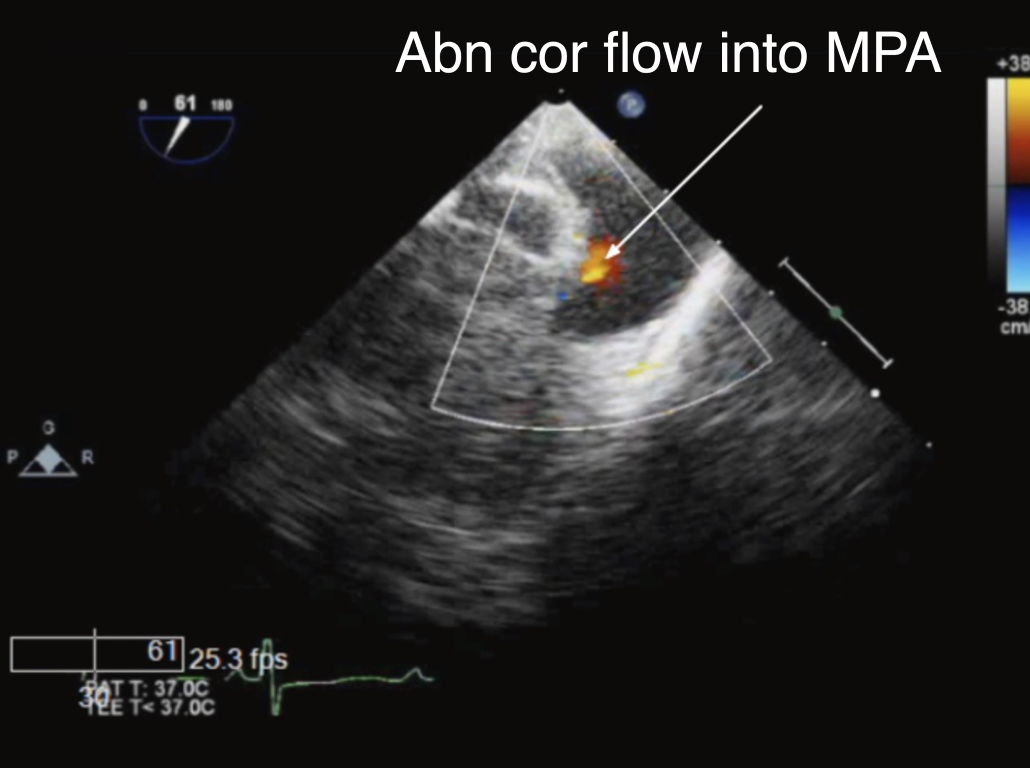
It remains to date not known if this method is applicable in patients with pulmonary arterial hypertension PAH and abnormal aortic-to-pulmonary shunting. Flow between the stent and the vessel wall was. The use of Doppler imaging is an extension of B-mode US using the same basic physics of acoustic energy transmission.
Colour coded blood flow imaging is a recently introduced and commercially available technique encoding the rate of change of the backscatter echo of the blood cells into colour. What are the 14 steps of blood flow through the heart. Magnetic resonance imaging is also sometimes called nuclear.
The present case analyzes the effect of a patent ductus arteriosus PDA on pulmonary artery flow patterns in PAH mPAP from right heart catheterization 75 mmHg. The spectral analysis is typical displaying a pulsatile and positive waveform with blood flow velocity of 35 11 cmsec. Fourdimensional flow magnetic resonance imaging 4D flow MRI enables efficient investigation of cerebral blood flow pulsatility in the cerebral arteries.
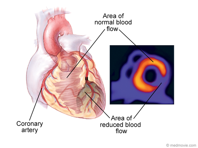
Depending on the angulations of the color box and the blood flow direction blood flow is displayed either in red or in blue. Blood flow rate and velocity in each stenosis segment were derived from the volumetric analysis of the FD-OCT pull back images. Those areas lacking blood flow demonstrate filling defects visualized between images taken at rest and under stress.
Abnormal flow dynamics such as vortex blood flow pattern in the pulmonary artery PA may reflect progression of pulmonary arterial hypertension PAH. It can be used to examine your heart and blood vessels and to identify areas of the brain affected by stroke. A contrast dye is injected into the patients arm and as the patient lies on a table the CT scanner rotates taking multiple images -- yielding highly detailed images of blood vessels and the heart.
A Doppler ultrasound can estimate how fast blood flows by measuring the rate of change in its pitch frequency. It can also show where blockages are and how severe they are. It concentrates in the areas of the heart that have the best blood flow.
Myocardial Perfusion Scan Resting Johns Hopkins Medicine

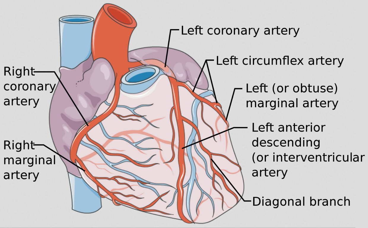
Cv Physiology Coronary Anatomy And Blood Flow
Thallium Stress Test
Diagnosis Of Myocardial Ischemia Radiology Key

Coronary Angiography Heart Test Recovery Risk Heart Foundation
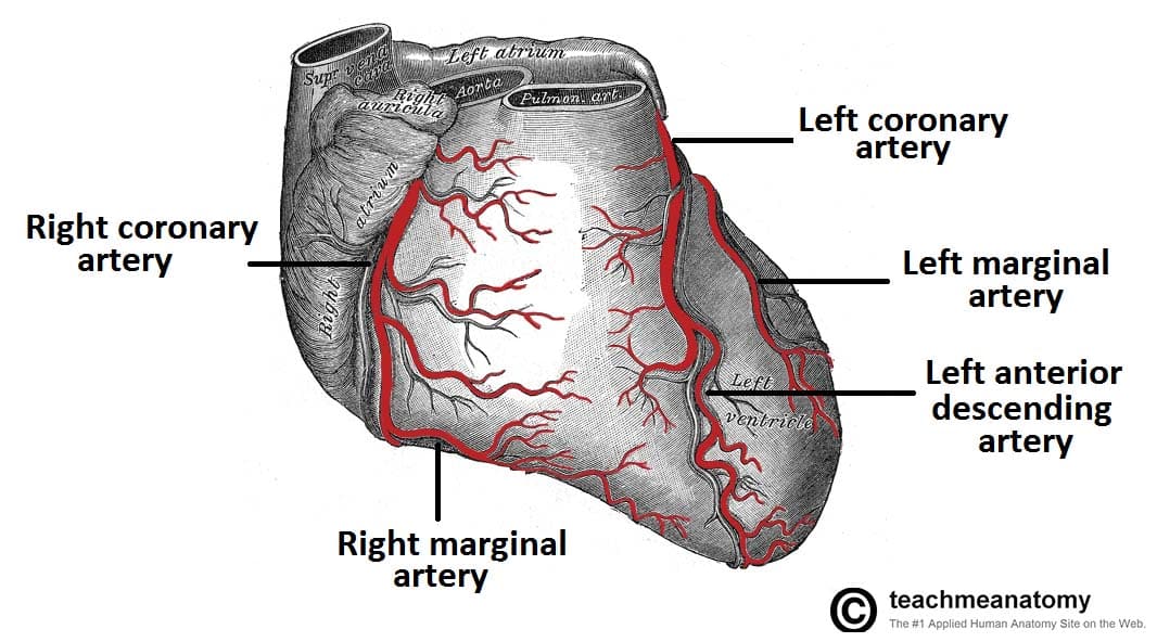
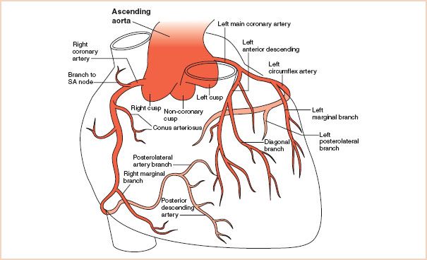
Vasculature Of The Heart Teachmeanatomy
Physiology Of Coronary Blood Flow Radiology Key
Asnc And Snmmi Issue Guide For Using Pet Myocardial Blood Flow To Improve Coronary Artery Disease Diagnosis Daic

These 3 Imaging Tests Can Help Determine Your Risk Of Stroke Rai Health Awareness Blog
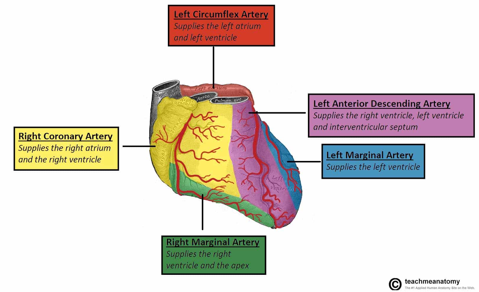
Vasculature Of The Heart Teachmeanatomy
Structure And Function Of Blood Vessels Anatomy And Physiology Ii

Artery Wall An Overview Sciencedirect Topics
Physiology Tutorial Coronary Circulation
Intraoperative Imaging Of The Coronary Arteries And Related Anomalies Congenital Cardiac Anesthesia Society
Intraoperative Imaging Of The Coronary Arteries And Related Anomalies Congenital Cardiac Anesthesia Society
Cv Physiology Coronary Anatomy And Blood Flow
Physiology Of Coronary Blood Flow Radiology Key

Coronary Arteries Radiology Reference Article Radiopaedia Org
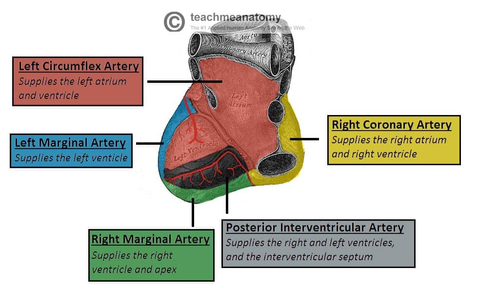
Vasculature Of The Heart Teachmeanatomy
0 Response to heart artery blood flow imaging
Posting Komentar